NYC’s Leading Integrative Health Care Center

Anti-Aging and Longevity Treatments Remember when you were in college and stayed up all night drinking beer, eating pizza, and partying; yet you still were able to attend class in the morning? How many of you could do that now?
Chronic bronchitis is a persistent inflammatory disease of the bronchial airways that traps excess mucus in the lungs, producing a daily productive cough that lasts for months and compounds into years of progressive breathing difficulty. For the millions living with this condition, each morning begins with an exhausting cough, and each flight of stairs becomes a negotiation with their own lungs.
Americans diagnosed with chronic bronchitis annually
Of cases linked to identifiable environmental triggers
Leading cause of death in the US (COPD-spectrum)
Average timeframe to first meaningful symptom relief with integrative care
Board-certified integrative medicine physician.
Chronic bronchitis is a form of chronic obstructive pulmonary disease (COPD) characterised by persistent inflammation and hypersecretion of mucus in the bronchial tubes, clinically defined as a productive cough lasting at least three consecutive months per year for two or more consecutive years in the absence of another identifiable cause. The inflammatory cascade—driven by inhaled irritants, oxidative stress, and immune dysregulation—leads to goblet cell hyperplasia, ciliary dysfunction, impaired mucociliary clearance, and progressive narrowing of the bronchial lumen. Functional medicine treats chronic bronchitis as a systemic inflammatory condition with identifiable, modifiable root causes rather than an irreversible structural disease, distinguishing it meaningfully from the conventional model of management alone.
Chronic bronchitis is a long-term disease of the airways in which the bronchial tubes—the branching passages that carry air from the trachea into the lungs—become persistently inflamed, swollen, and filled with excess mucus. Unlike a chest cold or acute bronchitis that resolves in two to three weeks, chronic bronchitis is a structural and immunological condition that becomes self-perpetuating: the inflammation promotes mucus overproduction, the excess mucus blocks airways and traps pathogens, and those pathogens trigger further inflammation in a cycle that can continue for decades without targeted intervention.
At the cellular level, chronic irritant exposure—most commonly tobacco smoke, but also occupational dust, diesel exhaust, coal, silica, and household air pollution—activates the bronchial epithelium’s inflammatory signalling cascade via nuclear factor kappa-B (NF-κB). This triggers recruitment of neutrophils and macrophages into the bronchial wall, release of proteases that break down connective tissue, and a compensatory hypertrophy of mucous glands that can increase in size by up to three times their normal volume (Reid index). The result is airways that are perpetually narrowed, poorly draining, and vulnerable to repeated bacterial colonisation by organisms such as Haemophilus influenzae, Streptococcus pneumoniae, and Moraxella catarrhalis.
Conventional medicine manages chronic bronchitis primarily through bronchodilators (short-acting beta-agonists such as albuterol and long-acting muscarinic antagonists), inhaled corticosteroids, and antibiotics during exacerbations—all of which address symptoms rather than underlying biology. Functional medicine adds a critical dimension: systematic investigation of the immunological, nutritional, environmental, and microbiological factors that determine why this patient’s airways remain chronically inflamed when a similar exposure in a different person does not produce the same response. Factors such as vitamin D deficiency, glutathione depletion, gut dysbiosis-driven systemic inflammation, heavy metal burden, and genetic polymorphisms in antioxidant enzymes (such as SOD2 and GSTM1) all influence bronchial inflammation susceptibility and are actionable targets for treatment.
Chronic bronchitis affects an estimated 9 million Americans aged 18 and older, with prevalence rising sharply in adults over 45. Women are diagnosed approximately 30% more often than men, a disparity thought to reflect differences in airway calibre, hormonal effects on bronchial reactivity, and greater susceptibility of smaller airways to inhaled irritants. The condition is also heavily correlated with socioeconomic disadvantage, occupational exposure, and urbanisation—factors that make an individualised functional medicine assessment particularly important for patients in high-exposure environments like New York City.
The bronchi and bronchioles are air-conducting tubes lined with ciliated epithelial cells and mucus-secreting goblet cells. In chronic bronchitis, the goblet cell population expands dramatically, goblet cells migrate into smaller airways where they are not normally present, and the cilia that sweep mucus out of the lung become damaged and dysfunctional.
Normally, mucus is produced in controlled amounts to trap and remove inhaled particles. In chronic bronchitis, the Reid index (ratio of mucous gland thickness to total airway wall thickness) exceeds 0.4, compared to the normal 0.26, flooding the airways with thick, purulent secretions that obstruct airflow and create a bacterial colonisation medium.
The mucociliary escalator is the lung’s primary self-cleaning mechanism, relying on coordinated ciliary beating to move mucus and trapped particles upward toward the throat for clearance. Chronic bronchitis destroys cilia through direct oxidative damage and protease activity, permanently impairing this defence and making mucolytic support—via NAC and adequate hydration—a critical treatment priority.
Chronic bronchitis produces a wide and often underappreciated spectrum of symptoms that extend well beyond the signature cough—encompassing energy, cognitive function, cardiovascular health, and immune resilience—because the underlying airway inflammation creates systemic inflammatory effects throughout the body.
The defining symptom: daily mucus-producing cough present on most days for at least 3 consecutive months, caused by goblet cell hyperplasia flooding the airways with excess secretions.
Initially on exertion only, then at rest as disease progresses; results from reduced airway lumen diameter caused by mucus, inflammation-induced wall oedema, and smooth muscle constriction.
High-pitched respiratory sounds produced by airflow through narrowed, mucus-clogged bronchioles; tightness reflects increased airway resistance against which respiratory muscles must work.
Recurrent bronchitis exacerbations caused by bacterial colonisation of stagnant mucus pools—each infection accelerates structural airway damage by triggering a fresh neutrophilic inflammatory surge.
Yellow, green, or grey sputum containing dead neutrophils and bacterial debris indicates active infection or colonisation by opportunistic pathogens between acute exacerbations.
Coughing is most severe on waking, as overnight mucociliary stagnation allows mucus to pool in the lower airways; nocturnal breathing in horizontal posture prevents the postural drainage that gravity provides during the day.
Persistent exhaustion driven by hypoxaemia reducing cellular ATP production, elevated catabolic cytokines (TNF-α, IL-1β) suppressing energy metabolism, and the extraordinary muscular effort of breathing against high airway resistance.
Nocturnal coughing episodes fragment sleep architecture, preventing restorative deep sleep and REM cycles; hypoxaemia during sleep further impairs overnight recovery and contributes to daytime cognitive dysfunction.
Lactic acid accumulates rapidly during exertion because hypoxic muscles shift to anaerobic metabolism; patients often experience breathlessness out of proportion to actual exertion, reinforcing sedentary behaviour.
Cerebral hypoperfusion from chronically reduced oxygen saturation (SpO₂ below 94%) impairs prefrontal cortex function, slowing processing speed, working memory, and executive decision-making.
Hypoxia-driven neuroinflammation and the chronic burden of a physically limiting respiratory disease create a high prevalence of comorbid depression (up to 25%) and anxiety (up to 37%) in chronic bronchitis patients.
Systemic pro-inflammatory cytokines from the bronchial inflammation cascade sensitise pain receptors throughout the body, creating widespread musculoskeletal discomfort that is frequently dismissed as a separate condition.
Fluid accumulation in the legs signals that pulmonary hypertension has begun placing excessive workload on the right ventricle (cor pulmonale), causing right-heart backpressure that forces fluid into peripheral tissues.
Bluish discolouration occurs when oxygen saturation falls below approximately 85%, causing deoxyhaemoglobin to dominate in peripheral capillaries; a clinical sign indicating moderate-to-severe impairment of gas exchange.
The cardiovascular system compensates for reduced oxygen delivery by increasing cardiac output; chronic tachycardia increases the risk of atrial arrhythmias and accelerates myocardial wear over time.
Advanced disease causes significant weight loss due to elevated resting energy expenditure from laboured breathing and catabolic cytokine activity; earlier disease may produce weight gain due to inactivity and systemic inflammation.
Chronic hypoxaemia stimulates erythropoietin production, increasing red blood cell mass in an attempt to enhance oxygen-carrying capacity; this thickens the blood and raises deep vein thrombosis and stroke risk.
In long-standing disease with air trapping, the thoracic cage gradually adopts a rounded, barrel-shaped appearance as the lungs remain in a semi-inflated state; ribs become more horizontal and the anteroposterior chest diameter increases.
Damaged bronchial epithelium, impaired mucociliary clearance, and immune exhaustion from chronic activation combine to create a substantially weakened first line of respiratory defence against bacteria and viruses.
Each acute exacerbation—triggered by new infection or irritant exposure—takes longer to resolve than in healthy individuals because the underlying airway architecture never fully returns to its pre-exacerbation state.
Chronic bronchial inflammation lowers the threshold for allergic responses throughout the respiratory tract; many patients develop new-onset allergic rhinitis, nasal polyps, or food sensitivities as the disease progresses.
The gut-lung axis connects bronchial inflammation to intestinal permeability and microbiome disruption; many patients experience bloating, irregular bowel habits, or reflux that worsens their respiratory symptoms by increasing intra-abdominal pressure against the diaphragm.
Clubbing of the fingertips—a bulbous enlargement of the fingernail bed—can develop in longstanding hypoxic disease; skin pallor and nail ridging reflect chronic nutritional depletion secondary to malabsorption and oxidative stress.
Chronic inflammation and poor appetite in advanced disease deplete zinc, selenium, magnesium, vitamins C, D, and E—all essential to bronchial immune defence; deficiency states amplify inflammation in a destructive feedback loop.
Chronic bronchitis severity is formally classified using the GOLD (Global Initiative for Chronic Obstructive Lung Disease) framework, which stratifies airflow limitation by the ratio of forced expiratory volume in one second to forced vital capacity (FEV1/FVC) on spirometry. Understanding which stage a patient is in guides treatment intensity, monitoring frequency, and realistic expectations for recovery—and at Patients Medical, we use staging not as a ceiling but as a baseline to measure how far patients improve.
Spirometry confirms an FEV1/FVC ratio below 0.70 but FEV1 remains at or above 80% of predicted. Most patients at this stage have a chronic cough and sputum production but rarely notice significant breathlessness. This is the most common stage at which chronic bronchitis goes undiagnosed because symptoms seem trivial and standard GP appointments are brief. Functional medicine intervention at this stage achieves the greatest long-term benefit: trigger elimination, antioxidant repletion, and anti-inflammatory nutrition can stabilise or reverse early airway changes before structural remodelling becomes entrenched.
FEV1 has declined to 50–79% of predicted, and dyspnoea on exertion becomes noticeable enough to prompt the majority of patients to seek medical care for the first time. Coughing is persistent and often disrupts sleep; exercise capacity is measurably reduced. At this stage, mucous gland hypertrophy is well established and bacterial colonisation of the lower airways (most commonly Haemophilus influenzae) is common even between acute exacerbations. Personalised pulmonary rehabilitation combined with IV antioxidant therapy and microbiome support can significantly improve symptom burden and reduce exacerbation frequency at this stage.
FEV1 is 30–49% of predicted and patients experience significant exercise intolerance, frequent acute exacerbations (two or more per year), and often require supplemental oxygen. Pulmonary hypertension may be developing, and right ventricular strain begins to appear on echocardiography. The quality-of-life impact is profound: many patients become housebound and struggle with depression and social isolation. Integrative treatment at this stage focuses on reducing exacerbation frequency through immune support, ozone and UV blood irradiation therapy, and rigorous nutritional optimisation, alongside conventional bronchodilator management.
FEV1 below 30% of predicted constitutes respiratory failure and is characterised by disabling breathlessness at rest, continuous oxygen dependence, and high risk of life-threatening exacerbations. Cor pulmonale with peripheral oedema is typically present. Patients at this stage require an integrated care team including pulmonology, integrative medicine, and cardiac monitoring. Functional medicine goals shift toward exacerbation prevention, optimising nutritional status to support respiratory muscle strength, and improving quality of life—goals that remain achievable and meaningful even at this advanced stage.
Chronic bronchitis almost never results from a single cause. In the vast majority of patients, the condition develops and persists because of a convergence of multiple inflammatory triggers—environmental, nutritional, immunological, and microbial—acting simultaneously on airways that are rendered susceptible by genetic, metabolic, and lifestyle vulnerabilities. Understanding this multiplicity is precisely why a root-cause functional medicine approach produces results that symptom management alone cannot.
The single largest risk factor, responsible for approximately 70–80% of cases; tobacco smoke contains over 4,000 chemicals that directly damage bronchial cilia, activate NF-κB inflammatory signalling, and deplete glutathione—the lung’s primary antioxidant—with each puff.
Long-term exposure to coal dust, silica, grain dust, welding fumes, isocyanates, and cadmium in workplaces causes an occupational form of chronic bronchitis that develops independently of smoking and accounts for up to 15% of all COPD cases.
Fine particulate matter (PM2.5) from vehicle exhaust, ozone, nitrogen dioxide, and indoor pollutants (cooking fumes, mould spores, volatile organic compounds) trigger the same bronchial inflammatory cascade as tobacco smoke in susceptible individuals.
Each viral or bacterial respiratory infection inflicts direct epithelial damage and leaves residual inflammatory programming; patients who experience three or more lower respiratory infections per year are at substantially elevated risk for developing persistent bronchial inflammation.
Vitamin D3 (calcitriol) is an essential regulator of bronchial innate immunity; serum levels below 30 ng/mL impair the production of antimicrobial peptides (cathelicidin, defensins), increase susceptibility to viral infections, and amplify the NF-κB inflammatory response in bronchial epithelial cells.
The lungs are exposed to the highest ambient oxygen levels of any organ and require abundant glutathione, superoxide dismutase, and catalase to neutralise reactive oxygen species; smoking, poor nutrition, ageing, and genetic polymorphisms in GSTM1 and SOD2 enzymes all reduce antioxidant reserves, accelerating bronchial oxidative damage.
The gut-lung axis is a bidirectional immunological network: intestinal dysbiosis with Bacteroidetes depletion and Proteobacteria overgrowth increases systemic LPS (lipopolysaccharide) translocation, elevating circulating inflammatory cytokines that prime the bronchial immune environment for chronic inflammation.
This genetic condition—caused by mutations in the SERPINA1 gene—reduces levels of the protease inhibitor alpha-1 antitrypsin in the lungs, allowing neutrophil elastase to destroy bronchial connective tissue unchecked; patients often develop severe COPD before age 45, even without significant smoke exposure.
Micro-aspiration of gastric acid into the airways during nocturnal reflux events directly damages bronchial epithelium and triggers localised inflammation; GERD is present in 30–50% of chronic bronchitis patients and significantly worsens disease severity when untreated.
Cadmium (from cigarette smoke and industrial exposure), lead, arsenic, and mercury accumulate in bronchial tissue, directly inhibit antioxidant enzyme activity, impair mucociliary function, and have been identified as independent risk factors for COPD-spectrum disease in multiple large epidemiological studies.
Chronic inhalation of Aspergillus, Stachybotrys, and Cladosporium mycotoxins from water-damaged buildings triggers persistent bronchial and alveolar inflammation in susceptible individuals; this cause is frequently missed without mould toxin testing and detailed environmental history.
Chronic psychosocial stress elevates cortisol and activates the sympathetic nervous system in ways that suppress regulatory T-cell function, reduce secretory IgA in the respiratory tract, and lower resistance to infection—mechanisms that explain the consistent epidemiological finding that poverty, stress, and social disadvantage independently increase COPD risk.
Because chronic bronchitis shares many symptoms—cough, breathlessness, mucus production—with several other respiratory conditions, accurate diagnosis requires careful differentiation. The table below compares chronic bronchitis with the conditions it is most commonly confused with or co-occurs alongside.
| Feature | Chronic Bronchitis | Asthma | Emphysema | Bronchiectasis |
|---|---|---|---|---|
| Key Mechanism | Mucous gland hyperplasia + goblet cell proliferation causing mucus hypersecretion | Airway hyperreactivity with episodic bronchospasm (eosinophilic/allergic) | Alveolar wall destruction and air sac enlargement reducing gas exchange surface | Permanent bronchial dilation from recurrent infection and inflammation destroying airway walls |
| Hallmark Symptom | Daily productive cough with mucus for ≥3 months/year, ≥2 consecutive years | Episodic wheeze, cough, and chest tightness — often worse at night | Progressive dyspnoea with minimal cough; often “pink puffer” appearance | Large volumes of purulent sputum, haemoptysis (coughing blood), recurrent pneumonia |
| Best Diagnostic Test | Spirometry (FEV1/FVC <0.70) + clinical history of productive cough duration | Spirometry with >12% bronchodilator reversibility + methacholine challenge | High-resolution CT (HRCT): centrilobular or panlobular emphysematous changes | High-resolution CT: cylindrical, varicose, or cystic bronchial dilation |
| Standard Blood Test Findings | Elevated hs-CRP, fibrinogen; neutrophilia; possible polycythaemia (raised haematocrit) | Raised IgE; eosinophilia; raised exhaled nitric oxide (FeNO); allergen-specific IgE | Normal or polycythaemia; low pO₂ and raised pCO₂ on arterial blood gas | Raised ESR/CRP; sputum culture positivity; Ig levels to exclude immune deficiency |
| Treatment Approach | Trigger elimination, mucolytics (NAC), bronchodilators, anti-inflammatory nutrition, IV antioxidant therapy | Inhaled corticosteroids + long-acting bronchodilators; allergen avoidance; immunotherapy | Smoking cessation (most critical); bronchodilators; supplemental oxygen; lung volume reduction | Airway clearance physiotherapy; targeted antibiotic courses; bronchodilators; surgery in select cases |
| Functional Medicine Overlap | Gut microbiome, heavy metals, nutritional status, mould exposure, GERD all addressable | Significant overlap: IgG food sensitivities, vitamin D, omega-3 status, gut-lung axis | Antioxidant and anti-inflammatory nutrition extends utility beyond cessation alone | Microbiome, immunoglobulin, and nutritional status all influence exacerbation frequency |
At Patients Medical, chronic bronchitis diagnosis begins with a thorough clinical history but extends far beyond what a standard GP visit or emergency evaluation covers. Our five-step diagnostic protocol is designed to establish not just the presence of chronic bronchitis but the specific biological drivers maintaining it in this individual patient—information that is essential to crafting a treatment plan that achieves lasting improvement rather than temporary symptom relief.
Spirometry measures FEV1 (the volume of air exhaled forcefully in one second) and FVC (total forced vital capacity). An FEV1/FVC ratio below 0.70 after inhaled bronchodilator administration confirms obstructive airflow limitation consistent with COPD-spectrum disease. This test definitively distinguishes chronic bronchitis from restrictive lung diseases and establishes the GOLD stage, which guides treatment intensity. We use body plethysmography for comprehensive lung volume measurement in complex cases.
Standard chest X-rays miss the early and moderate-stage airway changes that define chronic bronchitis. HRCT provides millimetre-resolution images of airway wall thickening, mucus plugging, bronchial dilation, and early emphysematous changes (centrilobular or panlobular). It also identifies complicating factors such as pulmonary nodules, bronchiectasis, or pleural disease that would alter management. We routinely use low-dose CT protocols to minimise radiation exposure while preserving diagnostic accuracy.
We measure high-sensitivity C-reactive protein (hs-CRP), interleukin-6 (IL-6), tumour necrosis factor-alpha (TNF-α), fibrinogen, complete blood count with differential, serum immunoglobulins (IgA, IgG, IgM, IgE), and total white cell count to characterise the inflammatory phenotype (neutrophilic vs eosinophilic), identify immune deficiency states, and establish baseline biomarkers for monitoring treatment response over time.
Sputum culture identifies the specific bacterial colonisers present in the airways—most commonly Haemophilus influenzae, Streptococcus pneumoniae, Moraxella catarrhalis, or Pseudomonas aeruginosa in severe disease—and their antimicrobial sensitivities, allowing targeted rather than empirical antibiotic selection during exacerbations. We also assess gut microbiome composition via our comprehensive stool testing because intestinal dysbiosis is a frequently missed systemic driver of bronchial inflammation through the gut-lung axis.
Our integrative evaluation includes comprehensive allergy testing (inhalant allergens: dust mites, mould, pet dander, pollen; food sensitivities via IgG4 panel), vitamin D3 and vitamin C serum levels, heavy metal testing including urinary cadmium (the most relevant lung toxin), zinc, magnesium, and selenium status, mould mycotoxin testing for indoor environmental exposure, and alpha-1 antitrypsin genotyping for patients under 50 or without significant smoking history. This panel routinely identifies actionable deficiencies or exposures missed by standard evaluations.
Check all that apply to your current experience:
Our anxiety treatment protocol is personalised to your laboratory results — not a one-size-fits-all approach. We address the specific biochemical drivers identified in your testing workup, using interventions with the strongest evidence and the least side-effect burden.
The single most effective intervention in chronic bronchitis is the sustained removal of the inflammatory trigger maintaining the condition. At Patients Medical, we conduct a systematic environmental and occupational history combined with objective testing to identify all active triggers: residual tobacco exposure, occupational inhalants, indoor mould and mycotoxins, GERD, and unrecognised allergens. Elimination is guided, supported, and monitored—because patients who have lived with their triggers for years often need both practical strategies and biological support to make sustained change.
Evidence-based nutraceutical protocols form the foundation of our anti-inflammatory bronchial repair programme. N-acetylcysteine (NAC, 600–1200mg daily) both replenishes glutathione and reduces mucus viscosity through direct mucolytic activity. Vitamin D3 (dosed to achieve 60–80 ng/mL serum) restores antimicrobial peptide production in the bronchial epithelium. Quercetin (1000mg daily) inhibits mast cell degranulation and NF-κB signalling. Omega-3 fatty acids (EPA/DHA, 3–4g daily) shift the arachidonic acid cascade toward less inflammatory leukotrienes. Zinc and selenium support innate immune function and ciliary structure. All protocols are dosed based on individual testing.
Intravenous delivery bypasses the gastrointestinal tract to achieve plasma concentrations of key antioxidants that are unattainable orally. Our bronchitis IV protocols include high-dose vitamin C (10–50g per infusion), reduced glutathione (600–1200mg), zinc, magnesium, B-complex vitamins, and selenium—nutrients that directly neutralise reactive oxygen species in the bronchial epithelium, reduce pro-inflammatory cytokine production, and accelerate mucosal repair. Most patients notice reduced cough frequency and improved energy within 3–6 infusions.
Medical ozone therapy (ozone autohemotherapy) and ultraviolet blood irradiation (UBI) are advanced integrative modalities used at Patients Medical for patients with chronic respiratory infections, bacterial colonisation, and immune dysregulation driving their bronchitis. Ozone activates the nuclear factor erythroid 2-related factor 2 (Nrf2) pathway, upregulating the body’s own antioxidant production (glutathione, superoxide dismutase, catalase) while exerting direct antimicrobial effects. UBI improves lymphocyte activity and has been shown in clinical practice to reduce the frequency of respiratory infections in COPD patients.
Exercise-based pulmonary rehabilitation is one of the highest-evidence interventions for COPD-spectrum disease: systematic review data shows it reduces hospitalisation rates by 40% and significantly improves quality of life and exercise capacity regardless of disease severity. We coordinate supervised pulmonary rehabilitation programmes tailored to each patient’s current FEV1 and exercise tolerance, combined with breathing retraining techniques—including diaphragmatic breathing, pursed-lip breathing, and the Buteyko method for CO₂ tolerance—that reduce the work of breathing, calm respiratory anxiety, and improve functional capacity for daily activities.
Restoring gut microbiome diversity is a cornerstone of long-term bronchial inflammation control via the gut-lung axis. Our approach combines comprehensive stool testing to identify dysbiosis patterns, targeted probiotic therapy (especially Lactobacillus rhamnosus GG, Bifidobacterium longum, and Saccharomyces boulardii), prebiotic dietary protocols, and leaky gut repair using L-glutamine, zinc carnosine, and deglycyrrhizinated liquorice (DGL). This is combined with a personalised anti-inflammatory nutrition plan that eliminates identified food sensitivities and emphasises Mediterranean-style eating patterns that reduce systemic TNF-α and IL-6 levels, reducing the inflammatory load on the bronchial mucosa.
| Weeks 1–6 | Comprehensive testing completed; primary trigger identified and eliminated; IV antioxidant series initiated; NAC and vitamin D protocols started. Most patients notice reduced cough frequency and improved morning symptoms within 3–4 weeks. |
| Months 2–6 | Nutraceutical protocols optimised based on initial response; pulmonary rehabilitation programme underway; gut microbiome restoration advancing; serial hs-CRP and sputum culture monitoring to confirm reduction in inflammatory load. FEV1 testing at 3 months establishes objective baseline trajectory. |
| Months 6–18 | Ongoing monitoring with annual spirometry; maintenance nutraceutical protocol; dietary and lifestyle optimisation; exacerbation prevention plan. Patients who achieve full trigger elimination and sustain nutritional optimisation consistently maintain the improvements achieved during active treatment. |
Targeted lifestyle changes are not optional add-ons to chronic bronchitis treatment—they are primary therapeutic interventions that directly modulate bronchial inflammation, mucociliary function, and immune competence. The following practices are specific, evidence-supported actions rather than generic wellness advice.

Perform 15 minutes of active cycle of breathing technique (ACBT) each morning: 5 repetitions of thoracic expansion exercises followed by 3–4 forced expirations (huffs) at mid-lung volume, then a full cough. This is more effective than unstructured coughing at mobilising and clearing secretions while minimising airway collapse. Add gravity-assisted postural drainage (lying head-down at 20–30 degrees) for three minutes on each lateral side to enhance mucus movement from the lower lobes.

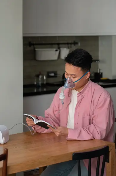
Adequate systemic hydration is the cheapest and most underutilised mucolytic agent available: bronchial secretions are 97% water, and even mild dehydration (urine osmolality above 800 mOsm/kg) measurably increases mucus viscosity and impairs mucociliary clearance. Target a minimum of 35ml/kg body weight in plain water daily (approximately 2.5L for a 70kg adult), warmed herbal teas (ginger, thyme, eucalyptus) in cooler months, and reduce diuretic beverages. Add a medical-grade saline nebuliser (0.9% or 3% hypertonic saline) for 10–15 minutes daily to hydrate airways from within.

Exercise is one of the strongest anti-inflammatory interventions available for chronic bronchitis, reducing TNF-α and IL-6 through PGC-1α-mediated mitochondrial biogenesis and skeletal muscle release of anti-inflammatory myokines (IL-6, IL-10, brain-derived neurotrophic factor). Begin with 20 minutes of level walking 3 days per week at a pace that permits conversation; add 5 minutes per week until reaching 45 minutes daily. Pursed-lip breathing during exertion (exhale through slightly pursed lips over 4–6 counts) reduces dynamic air trapping, slows respiratory rate, and allows higher exercise intensities to be sustained comfortably.

Chronic breathlessness activates the sympathetic nervous system in a feedback loop that worsens bronchospasm and anxiety. Break this cycle with 10 minutes of daily 4-7-8 breathing (inhale for 4 counts, hold for 7, exhale for 8), performed in a reclined position with hands on the abdomen. This activates the vagus nerve, reduces cortisol, lowers bronchial smooth muscle tone, and has been shown in small trials to reduce rescue inhaler use in COPD patients within two weeks of consistent practice. Consistency matters more than duration: 10 minutes daily outperforms 60 minutes occasionally.

NYC apartments frequently harbour mould, dust mites, volatile organic compounds (VOCs) from new furniture and cleaning products, and high particulate matter from cooking and outdoor ingress. Install a high-efficiency HEPA air purifier (minimum 350 CFM capacity) in the bedroom and keep windows closed during high-pollution days (AQI above 100). Replace household cleaning products with white vinegar, baking soda, and plant-based alternatives. Maintain indoor humidity at 45–55% to inhibit mould growth. Use a bedroom HEPA vacuum weekly with a sealed bag to remove dust mite antigen from soft furnishings—dust mites are present in 100% of New York City apartments.

Sleeping flat promotes mucus pooling in dependent lung segments and worsens nocturnal coughing and oxygen desaturation. Elevate the head of the bed by 30–45 degrees using a firm wedge pillow (not stacked standard pillows, which collapse under body weight) to keep airways open and facilitate continuous mucociliary drainage throughout the night. If nocturnal oxygen saturation monitoring shows SpO₂ dips below 90%, discuss overnight supplemental oxygen with your physician; even brief desaturations accelerate pulmonary vascular disease. Establish a fixed sleep schedule of 7–8 hours to support immune regulatory function—interleukin-10 (IL-10) production peaks during slow-wave sleep and plays a key role in bronchial anti-inflammatory regulation.
Diet is a primary determinant of systemic inflammatory tone in chronic bronchitis: the foods consumed daily either amplify or dampen the NF-κB-driven bronchial inflammation that perpetuates mucus hypersecretion, airway remodelling, and susceptibility to infection. A Mediterranean-style, nutrient-dense dietary pattern consistently demonstrates the greatest anti-inflammatory benefit in respiratory disease research, and it is the framework we adapt to each patient’s individual needs and sensitivities at Patients Medical.
Eliminate all processed, ultra-processed, and refined-carbohydrate foods completely for a minimum of 8 weeks. These foods are the primary dietary drivers of systemic NF-κB activation—the central inflammatory switch in chronic bronchitis—and their removal consistently produces a measurable reduction in hs-CRP and sputum production within 3–4 weeks, a change that no supplement can replicate while these foods remain in the diet.
Chronic bronchitis rarely exists in isolation. The same inflammatory mechanisms—NF-κB activation, oxidative stress, immune dysregulation—that drive bronchial mucus hypersecretion also affect other organ systems, making these comorbidities common, clinically important, and often simultaneously improvable through integrative care.
Chronic bronchitis is the dominant form of COPD by prevalence; when accompanied by emphysematous destruction of alveolar tissue, the two diseases combine to produce severe, compounded airflow limitation. Understanding both components is essential for accurate staging and treatment. Our full COPD guide addresses the emphysema component in detail.
Asthma and chronic bronchitis overlap in Asthma-COPD Overlap Syndrome (ACOS), where patients have features of both—eosinophilic airway inflammation, allergic sensitisation, and partially reversible obstruction alongside chronic mucus hypersecretion. ACOS responds to a distinct combined treatment approach addressing both allergic hyperreactivity and chronic structural airway changes.
Increased intestinal permeability allows bacterial lipopolysaccharide (LPS) and undigested food antigens to enter systemic circulation, triggering a chronic low-grade inflammatory state that primes bronchial immune cells for exaggerated responses to inhaled irritants. Treating leaky gut is frequently one of the highest-yield interventions for patients with treatment-resistant bronchitis.
The systemic hypoxaemia, mitochondrial dysfunction, and chronic cytokine elevation of chronic bronchitis create an overlap with chronic fatigue syndrome in many patients, producing fatigue that persists beyond what spirometry alone would predict. Addressing both the respiratory and metabolic components simultaneously achieves better energy recovery.
Inhalant allergies to dust mites, mould, animal dander, and seasonal pollens are among the most common and most overlooked triggers for chronic bronchitis exacerbations. Identifying and managing specific allergen sensitivities—rather than relying on non-specific antihistamines—reduces the bronchial inflammatory burden at its source.
Chronic bronchitis accelerates cardiovascular disease through pulmonary hypertension, systemic inflammation, hypoxaemia-driven polycythaemia, and shared risk factors (smoking, sedentary behaviour, oxidative stress). Patients with both conditions require coordinated cardiovascular and pulmonary assessment to avoid under-treating either component. Our cardiac testing panel includes NT-proBNP and echocardiography referral coordination.
Many people with chronic bronchitis spend years managing symptoms on their own—or with only sporadic antibiotic courses—without ever receiving a comprehensive evaluation of what is driving their condition. If any of the scenarios below apply to you, a thorough functional medicine assessment has the potential to identify root causes and treatment opportunities that have been missed.
🚨 Seek Emergency Medical Evaluation Immediately if You Experience:
The following testimonials represent the real experiences of patients who came to Patients Medical seeking answers after years of managing chronic respiratory symptoms. Names are anonymised to protect privacy. Individual results vary; these accounts do not constitute a guarantee of outcomes.
Chronic bronchitis is a long-term inflammatory disease of the bronchial tubes defined by a productive (mucus-producing) cough present on most days for at least three consecutive months per year, for two or more consecutive years. This distinguishes it from acute bronchitis, which is a short-term infection (usually viral) that resolves within two to three weeks without lasting airway changes.
In chronic bronchitis, the bronchial walls undergo structural remodelling: the mucus-secreting goblet cells multiply and migrate into smaller airways where they are not normally found, mucous glands hypertrophy (the Reid index—the ratio of gland thickness to airway wall thickness—rises from a normal 0.26 to above 0.4), and the cilia responsible for clearing mucus become damaged and dysfunctional. This creates a self-perpetuating cycle of mucus accumulation, bacterial colonisation, and renewed inflammation.
Functionally, chronic bronchitis is classified as a form of COPD when accompanied by persistent airflow limitation on spirometry (FEV1/FVC ratio below 0.70 after bronchodilator). Functional medicine approaches it as a systemic inflammatory condition with multiple identifiable and modifiable root causes, rather than simply a structural consequence of past smoke exposure.
Yes, chronic bronchitis can meaningfully improve with targeted treatment — and the improvement timeline depends primarily on the severity of structural airway changes present and how completely the underlying triggers can be eliminated. Most patients notice a reduction in morning mucus production and cough frequency within 4–8 weeks of trigger elimination combined with NAC and IV antioxidant protocols. Meaningful improvement in exercise tolerance and energy levels typically follows within 8–16 weeks.
Spirometry improvement — measurable increases in FEV1 — usually becomes detectable at the 3–6 month mark. Research consistently shows that sustained trigger elimination (especially smoking cessation) combined with pulmonary rehabilitation reduces the annual rate of FEV1 decline by 50% or more and substantially reduces exacerbation frequency. Structural changes to the bronchial walls that have accumulated over many years cannot always be fully reversed, but disease progression can be halted and symptoms reduced to manageable levels in nearly all patients.
The full integrative programme at Patients Medical typically runs 12–18 months of active treatment before transitioning to long-term maintenance monitoring. Patients who complete the full programme report the most sustained outcomes, because by that point the root causes have been addressed systematically rather than episodically.
The gold-standard diagnostic tests for chronic bronchitis are spirometry with bronchodilator reversibility (confirming an FEV1/FVC ratio below 0.70 consistent with obstructive airflow limitation) and a thorough clinical history documenting daily productive cough duration. High-resolution chest CT scan provides detailed airway imaging that standard X-rays miss entirely — including airway wall thickening, mucus plugging, and early emphysematous changes.
What standard evaluations routinely miss is the array of root-cause drivers maintaining the inflammation. A comprehensive functional medicine workup adds: high-sensitivity CRP and cytokine panel (to characterise the inflammatory phenotype), sputum culture (to identify bacterial colonisers), serum vitamin D, zinc, selenium and glutathione assessment, comprehensive allergy panel (IgE and IgG food and environmental), heavy metal testing including urinary cadmium, mould mycotoxin panel for indoor exposure, gut microbiome assessment via comprehensive stool testing, and alpha-1 antitrypsin genotyping in younger patients.
This additional testing routinely identifies actionable causes — mould exposure, severe nutritional deficiency, bacterial colonisation patterns, gut dysbiosis — that standard pneumology appointments simply do not have the time or scope to investigate. These findings directly shape the personalised treatment plan and are what differentiate an integrative approach from a symptom-management one.
Yes — profound fatigue is one of the most debilitating and frequently dismissed symptoms of chronic bronchitis, and it operates through several distinct mechanisms that compound one another. The most direct cause is hypoxaemia: when thickened, mucus-filled airways cannot efficiently oxygenate the blood, every cell in the body receives less oxygen than it needs for normal ATP production. The mitochondria cannot meet energy demands, resulting in a fatigue that no amount of sleep fully resolves.
Second, chronically elevated inflammatory cytokines — particularly TNF-α, IL-1β, and IL-6 — directly suppress energy metabolism, inhibit appetite, and create a ‘sickness behaviour’ state characterised by exhaustion, low mood, and cognitive slowing. This cytokine-driven fatigue is the same mechanism responsible for the profound fatigue of influenza or major surgery, sustained chronically at a lower intensity. Third, the greatly increased work of breathing in obstructed airways burns significantly more calories than normal respiration, leaving less metabolic energy for other activities. Sleep disruption from nocturnal coughing creates a fourth layer of cumulative energy deficit.
Addressing fatigue effectively in chronic bronchitis requires treating all four mechanisms simultaneously: improving gas exchange through airway treatment and bronchodilation, reducing systemic cytokine levels through anti-inflammatory nutrition and IV antioxidant therapy, optimising nutritional status (especially iron, vitamin B12, coenzyme Q10, and vitamin D, which are commonly depleted), and achieving consistent restorative sleep through nocturnal airway optimisation.
Chronic bronchitis and asthma are both airway inflammatory diseases, but they differ fundamentally in their mechanism, patient profile, reversibility, and natural history. Asthma is primarily a hyperreactive airway disease driven by allergen-specific or non-specific triggers (cold air, exercise, stress); its hallmark is episodic, largely reversible bronchospasm with more than 12% improvement in FEV1 following bronchodilator administration. Airway inflammation in asthma is eosinophilic, IgE-mediated, and allergically driven.
Chronic bronchitis involves structural goblet cell and mucous gland changes driven by chronic irritant exposure (most commonly tobacco smoke), producing persistent mucus hypersecretion that is present daily regardless of trigger exposure. Airway obstruction is neutrophilic and only partially reversible. Chronic bronchitis most commonly develops after age 40 in people with significant smoke or occupational inhalant exposure; asthma typically presents earlier, often in childhood, with a strong atopic background (eczema, allergic rhinitis, elevated IgE).
Some patients have features of both — a condition now termed Asthma-COPD Overlap Syndrome (ACOS) — which requires a distinct management approach. The simplest clinical differentiator is the daily productive cough: a persistent productive cough present on most days regardless of season or trigger is a hallmark of chronic bronchitis, not asthma.
Chronic bronchitis exerts significant cardiovascular strain through several pathways. The most direct is pulmonary hypertension: when chronic hypoxaemia causes the pulmonary arteries to constrict (hypoxic vasoconstriction), the right ventricle must work against increased resistance, eventually developing cor pulmonale — right-sided heart failure characterised by ankle oedema, jugular venous distension, and worsening breathlessness. Ankle swelling in a bronchitis patient is an important clinical sign that warrants urgent evaluation for right heart strain.
Simultaneously, the chronic systemic inflammation of bronchitis — evidenced by elevated hs-CRP, fibrinogen, and IL-6 — accelerates endothelial dysfunction and atherosclerosis, increasing cardiovascular event risk independent of other risk factors. Studies show patients with COPD-spectrum disease have a 2–3 fold higher risk of myocardial infarction and stroke compared to matched controls. Hypoxaemia also drives polycythaemia (excess red blood cell production), thickening the blood and elevating deep vein thrombosis and pulmonary embolism risk.
A comprehensive functional medicine evaluation for chronic bronchitis therefore includes cardiac risk assessment: NT-proBNP for early right heart strain, hs-CRP, omega-3 index, and lipid particle size testing. Antioxidant therapy, omega-3 optimisation, and anti-inflammatory nutrition address these cardiovascular co-risks simultaneously with the respiratory disease.
Several evidence-supported nutritional agents can meaningfully reduce bronchial inflammation and mucus production in chronic bronchitis. N-acetylcysteine (NAC) at 600–1200mg daily is the most extensively studied: it replenishes glutathione (the lung’s primary antioxidant), acts as a direct mucolytic by breaking disulphide bonds in mucus proteins, and has demonstrated reduced exacerbation frequency in multiple randomised controlled trials. It is a core component of every Patients Medical bronchitis protocol.
Vitamin D3 (dosed to achieve serum levels of 60–80 ng/mL, typically 5,000–10,000 IU daily) modulates both innate and adaptive bronchial immunity, upregulating cathelicidin and defensin antimicrobial peptide production and reducing respiratory infection frequency. Quercetin (500–1,000mg daily) inhibits NF-κB and mast cell degranulation. Magnesium glycinate (400–600mg daily) supports bronchial smooth muscle relaxation and reduces airway reactivity. Omega-3 fatty acids (EPA/DHA, 3–4g daily) shift eicosanoid production toward less inflammatory leukotrienes in the bronchial mucosa.
For patients with confirmed bacterial colonisation, berberine, oregano oil, and monolaurin can reduce pathogenic bacterial load without the antibiotic resistance risks of repeated pharmaceutical courses. All supplementation at Patients Medical is guided by individual baseline testing, as appropriate doses vary substantially based on absorption, genetic enzyme variants, and co-existing conditions. Self-prescribing without testing risks both under-dosing (ineffective) and over-dosing (harmful).
Patients Medical brings the depth of functional medicine investigation to chronic bronchitis — moving beyond inhaler prescriptions to identify and address the root causes that keep your airways inflamed, year after year. Our physicians treat the whole person, not just the spirometry number.
Call us at (212) 794-8800 · 800 Second Avenue, Suite 900, New York, NY 10017
Patients Medical specializes in gently helping the patient identify the root cause of their medical issues and then assist them to recover from their problems to help them move forward to good health.
To schedule an in person on Tele-medicine appointment, please call our office at (212) 794-8800 or email us at info@PatientsMedical.com We look forward to hearing from you
Patients Medical PC
1148 Fifth Avenue, Suite 1B New York, NY 10128
Copyright © 2025 Patients Medical. All Rights Reserved.
All information presented in this website is intended for informational purposes only and not for the purpose of rendering medical advice. Statements made on this website have not been evaluated by the Food and Drug Administration. The information contained herein is not intended to diagnose, treat, cure or prevent any disease. Patients Medical.