NYC’s Leading Integrative Health Care Center

Anti-Aging and Longevity Treatments Remember when you were in college and stayed up all night drinking beer, eating pizza, and partying; yet you still were able to attend class in the morning? How many of you could do that now?
Endometriosis occurs when tissue similar to the uterine lining implants outside the uterus and bleeds monthly with nowhere to go, triggering chronic inflammation, scar tissue, and pain. For many women, this means years of being told their pain is “just bad periods” before anyone investigates the underlying hormonal and inflammatory drivers.
women of reproductive age affected
average time to diagnosis
of infertility cases involve endometriosis
of disease classification
Board-certified integrative medicine physician.
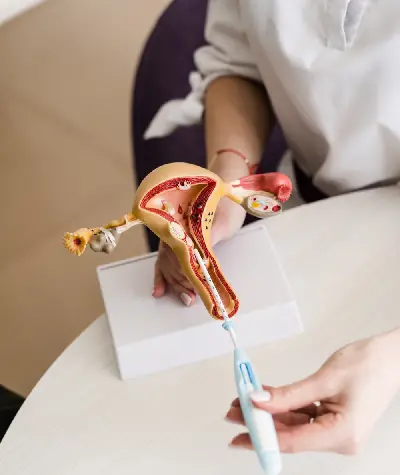
Endometriosis is a chronic inflammatory condition in which tissue similar to the endometrium grows outside the uterine cavity, commonly on the ovaries, fallopian tubes, and pelvic peritoneum. This ectopic tissue continues to respond to hormonal cycling, bleeding monthly without a natural exit, which drives local inflammation, adhesions, and often significant pelvic pain and infertility.
Endometriosis is a disease in which tissue that behaves like the uterine lining grows in places it should not, most commonly the ovaries, fallopian tubes, and the lining of the pelvis. Each menstrual cycle, this misplaced tissue thickens, breaks down, and bleeds in response to estrogen and progesterone signals, just as the normal uterine lining does. The critical difference is that this blood has no way to leave the body, so it pools locally, irritating surrounding tissue and triggering an ongoing inflammatory response.
At the biological level, three processes converge: hormone-driven tissue growth, an immune system that fails to clear the misplaced cells efficiently, and progressive fibrosis as the body attempts to wall off the irritation with scar tissue and adhesions. Over time, adhesions can bind the ovaries, fallopian tubes, bowel, and bladder together, distorting pelvic anatomy and contributing to both pain and infertility.
Conventional gynecology recognizes endometriosis as a surgically diagnosed structural disease, confirmed by direct visualization of lesions during laparoscopy. Functional medicine works alongside that framework but places equal emphasis on why the tissue is behaving this way in a given patient, examining estrogen metabolism, gut-driven estrogen recirculation, and systemic inflammation as modifiable drivers that influence symptom severity even when surgery is also part of the plan.
Endometriosis affects an estimated 1 in 10 women of reproductive age globally, though prevalence may be underreported given how often symptoms are dismissed as normal menstrual pain. It is most commonly diagnosed between ages 25 and 35, though symptoms frequently begin in the teenage years, and it can also affect women approaching menopause.
Endometrial implants on the ovaries can form endometriomas, sometimes called “chocolate cysts,” which are fluid-filled sacs containing old, darkened blood. These cysts can reduce ovarian reserve over time.
The thin membrane lining the pelvic cavity is the most common site for superficial implants, which trigger localized inflammation and are a major contributor to chronic pelvic pain.
Adhesions can bind the fallopian tubes to nearby organs, distorting their normal position and interfering with the tube’s ability to capture and transport an egg toward the uterus.
Because endometrial implants can develop in many locations and drive both local inflammation and whole-body hormonal disruption, symptoms often extend well beyond the pelvis into energy, digestion, mood, and reproductive health.
Severe cramping caused by prostaglandins released from irritated implant tissue.
Ongoing discomfort from adhesions and nerve irritation, not limited to menstruation.
Pain during intercourse from deep implants or restricted pelvic mobility.
Often linked to coexisting adenomyosis or estrogen dominance.
Mid-cycle pain as an ovary with adhesions attempts to release an egg.
Visible bloating from inflammatory fluid retention and slowed motility.
From implants on or near the bowel wall, often worse during menstruation.
Cyclical bowel changes tied to prostaglandin surges.
Frequently reported during the days surrounding menstruation.
Secondary to gut dysbiosis common in endometriosis patients.
Driven by the metabolic cost of sustained systemic inflammation.
Inflammatory cytokines can affect concentration and mental clarity.
Both a response to chronic pain and a direct effect of hormonal fluctuation.
Pelvic pain and cortisol dysregulation both interfere with restful sleep.
Intensified by estrogen-progesterone imbalance.
From distorted pelvic anatomy or an inflamed reproductive environment.
Variable bleeding patterns tied to estrogen dominance.
Referred pain from pelvic nerve irritation.
Another downstream sign of estrogen-progesterone imbalance.
Seen when implants affect the bladder wall.
Staging matters because it helps predict surgical complexity and, to some degree, fertility impact, though stage does not always correlate directly with pain severity. Many women with minimal disease experience severe pain, while some with extensive disease report milder symptoms.
A few superficial implants, typically on the peritoneum or ovarian surface, with little to no scar tissue. Often found incidentally during fertility workups.
Common in younger women early in disease course
More numerous implants with deeper tissue penetration and mild adhesions. Pain is often more pronounced than in Stage I despite modestly increased lesion count.
Frequently diagnosed after years of dismissed symptoms
Multiple deep implants, small to medium endometriomas on one or both ovaries, and noticeable adhesions between pelvic structures.
Often associated with fertility challenges
Extensive deep implants, large endometriomas, and dense adhesions that can bind the ovaries, tubes, uterus, and bowel together, sometimes called a “frozen pelvis.”
Typically requires specialized excision surgery
No single cause explains every case of endometriosis; most patients have several interacting hormonal, immune, and environmental factors that together allow implants to establish and persist.
Menstrual blood flows backward through the fallopian tubes into the pelvis instead of leaving the body, depositing endometrial cells outside the uterus.
Elevated or poorly metabolized estrogen relative to progesterone fuels the growth of endometrial-type tissue wherever it implants.
A healthy immune system should clear stray endometrial cells; in endometriosis, this clearance process is inefficient, allowing implants to survive.
Having a first-degree relative with endometriosis raises individual risk roughly sevenfold, pointing to inherited susceptibility.
Certain gut bacteria patterns interfere with estrogen clearance via the enterohepatic pathway, increasing circulating estrogen available to feed implants.
Elevated inflammatory cytokines both result from and further stimulate the growth and persistence of endometrial lesions.
Endocrine-disrupting chemicals such as certain phthalates and dioxins have been associated with increased endometriosis risk in research studies.
Starting menstruation before age 11 increases lifetime estrogen exposure and total number of menstrual cycles, both correlated with higher risk.
Cycles shorter than 27 days mean more frequent retrograde menstruation events over a lifetime.
Some researchers believe certain cells outside the uterus can transform directly into endometrial-like tissue under hormonal influence.
Endometrial cells may travel through lymphatic or blood vessels to distant sites, explaining rare implants found outside the pelvis.
Elevated cortisol disrupts the balance between estrogen and progesterone and amplifies the inflammatory response to existing implants.
Endometriosis shares symptoms with several other gynecologic and pain conditions, which is a major reason diagnosis is so often delayed. Understanding the distinguishing features helps clarify next diagnostic steps.
| Feature | Endometriosis | Adenomyosis | Fibroids | Irritable Bowel Syndrome |
|---|---|---|---|---|
| Key biomarker | CA-125 (often elevated), estrogen metabolites | CA-125 (mildly elevated), thickened myometrium | Typically normal bloodwork | No specific biomarker; calprotectin to rule out inflammation |
| Best diagnostic test | Laparoscopy with biopsy | Pelvic MRI or transvaginal ultrasound | Transvaginal ultrasound or MRI | Symptom-based (Rome IV criteria), exclusion testing |
| Hallmark symptom | Cyclical pelvic pain worsening over time | Heavy, painful periods with an enlarged, tender uterus | Pelvic pressure, heavy bleeding, bulk symptoms | Abdominal pain tied to bowel habit changes, not menstrual cycle |
| Standard blood test detection | Not reliably detected by routine bloodwork | Not reliably detected by routine bloodwork | Not detected by bloodwork; found on imaging | Not detected; diagnosis of exclusion |
| Treatment approach | Hormone regulation, anti-inflammatory therapy, surgery | Hormone therapy, uterine-preserving procedures, hysterectomy in severe cases | Hormone therapy, myomectomy, uterine artery embolization | Dietary modification (e.g., low-FODMAP), gut microbiome support, stress management |
| Overlap with endometriosis | — | Coexists in roughly 1 in 5 endometriosis patients | Can coexist, both estrogen-sensitive conditions | Frequently misdiagnosed as IBS due to overlapping bowel symptoms |
Clinically important overlap: Adenomyosis and endometriosis are commonly confused because both cause painful, heavy periods, and roughly 20% of patients have both conditions simultaneously. If you have been told your imaging is “normal” but your pain persists, it’s worth being evaluated for both.
Measures estradiol, progesterone, and estrogen-to-progesterone ratio across the cycle to identify estrogen dominance, a key driver of implant growth that standard single-day bloodwork often misses.
A dried urine test that reveals how estrogen is being metabolized and cleared through the liver, identifying patterns linked to higher inflammatory estrogen metabolites that standard blood hormone panels cannot detect.
Non-invasive imaging that can identify ovarian endometriomas and, in experienced hands, some signs of deep infiltrating disease, helping guide whether surgical referral is warranted.
Evaluates systemic inflammation and the CA-125 tumor marker, which is frequently elevated in moderate to severe endometriosis and helps track treatment response over time.
Assesses gut bacteria populations involved in estrogen recirculation through the enterohepatic pathway, a root-cause factor rarely evaluated in a standard gynecology visit.
Check all that apply to your current experience:
There is no one-size-fits-all endometriosis protocol. Our physicians build a personalized plan around your specific hormone metabolites, inflammatory markers, and gut findings, aiming to reduce lesion activity and pain while supporting fertility goals where relevant.
Addresses estrogen dominance directly, using targeted nutraceuticals and, where appropriate, bioidentical progesterone support to rebalance the estrogen-to-progesterone ratio driving implant growth.
A structured dietary protocol that reduces prostaglandin-driven inflammation, easing pain intensity and supporting the body’s own ability to manage ectopic tissue.
Delivers high-dose vitamins, minerals, and antioxidants directly into the bloodstream to support tissue repair and reduce oxidative stress associated with endometrial lesions.
Targets the bacterial populations responsible for estrogen recirculation, helping the body clear excess estrogen rather than reabsorbing it through the gut lining.
Manual therapy techniques that release pelvic floor tension and adhesion-related restriction, often reducing pain and improving mobility alongside medical treatment.
For appropriate candidates, bioidentical progesterone can help counterbalance estrogen dominance and stabilize cycle-related symptom flares under close physician supervision.
What to expect: Most patients report reduced pain intensity within 8–12 weeks of starting a personalized protocol. More significant shifts in cycle regularity, inflammatory markers, and fertility indicators typically emerge over 3–6 months. We re-test key biomarkers, including estrogen metabolites and inflammatory markers, every 90 days to confirm the plan is working and adjust as needed.

Practice 10 minutes of slow, extended-exhale breathing (inhale for 4 counts, exhale for 8) each morning. This shifts your nervous system out of the stress-dominant state that raises cortisol and worsens estrogen-progesterone imbalance.

On days with significant pelvic pain, replace vigorous exercise with a 20-minute walk or gentle yoga flow targeting hip openers, which improves pelvic blood flow without adding inflammatory load from high-intensity training.

Apply a heating pad to the lower abdomen for 15–20 minutes during flares. Heat relaxes uterine and pelvic smooth muscle and can reduce prostaglandin-driven cramping intensity within minutes.

Go to bed and wake at the same time daily, even on weekends, to support healthy cortisol rhythm. Disrupted sleep timing raises next-day inflammatory markers, which can intensify pelvic pain.

Switch to glass or stainless steel food storage, avoid heating food in plastic, and choose fragrance-free personal care products to lower exposure to endocrine-disrupting chemicals linked to estrogen dominance.

Log pain level, bleeding, and digestive symptoms daily for at least two full cycles. This pattern data helps your physician correlate symptom flares with specific hormonal phases and fine-tune treatment timing.
Diet matters mechanistically because certain foods directly raise prostaglandin production and inflammatory signaling, while others support the liver and gut pathways responsible for clearing excess estrogen from circulation.
Reduce inflammatory omega-6-heavy vegetable oils (corn, soybean, sunflower) and increase omega-3 sources (fatty fish, flaxseed, walnuts) to shift your prostaglandin balance away from the pain-amplifying pathway.
These conditions frequently coexist with or mimic endometriosis, and screening for them can clarify a stalled or incomplete diagnosis.
Endometrial-type tissue growing into the uterine muscle wall itself, causing similar pain and heavy bleeding; present in roughly 1 in 5 endometriosis patients.
A hormonal condition involving irregular ovulation and elevated androgens that can coexist with endometriosis and complicate hormone testing interpretation.
Endometriosis is found in 30–50% of infertility cases, making joint evaluation of both conditions essential for family planning.
Bowel symptoms from endometriosis implants near the intestines are frequently misdiagnosed as IBS due to overlapping bloating and pain patterns.
A bladder pain condition that can coexist with endometriosis, sometimes called the “pelvic pain twins” due to overlapping urinary and pelvic symptoms.
Broader estrogen-progesterone disruption often underlies both endometriosis and other cycle-related symptoms across the reproductive years.
Many women wait years before seeking evaluation because their pain has been normalized. If any of the following apply to you, an evaluation is worth pursuing sooner rather than later.
🚨 Seek Emergency Medical Evaluation Immediately If: You experience sudden, severe abdominal pain accompanied by fever, fainting, or heavy bleeding that soaks through protection every hour, as these can signal a ruptured ovarian cyst, ovarian torsion, or another acute surgical emergency requiring urgent care rather than a scheduled visit.
The experiences below are representative patient scenarios reflecting the kind of outcomes patients pursuing this type of care may report; individual results vary.
Yes. Endometriosis is a well-documented, universally recognized chronic inflammatory disease affecting an estimated 1 in 10 women of reproductive age worldwide. It occurs when tissue similar to the endometrium implants and grows outside the uterine cavity, commonly on the ovaries, fallopian tubes, and pelvic peritoneum. Because this tissue has no exit route when it bleeds monthly, it triggers local inflammation, scar tissue, and adhesions. Diagnosis has historically taken an average of 7 to 10 years from symptom onset, largely because pain is often dismissed as normal menstrual cramping. Confirmatory diagnosis is made through laparoscopic visualization with tissue biopsy, though imaging, symptom history, and hormone testing can support a strong clinical suspicion before surgery. At Patients Medical, we combine conventional diagnostic recognition with a functional medicine approach that investigates the hormonal and inflammatory drivers behind each patient’s presentation.
There is no single timeline, because endometriosis is managed chronically rather than cured in weeks. Most patients notice initial improvements in pain and cycle regularity within 8 to 12 weeks of starting a personalized protocol addressing estrogen metabolism, inflammation, and gut health. More significant shifts, such as reduced reliance on pain medication or improved fertility markers, typically take 3 to 6 months of consistent treatment. Patients with more extensive disease or coexisting conditions like adenomyosis may need 6 to 12 months of combined intervention. Because endometriosis can recur even after surgical removal of lesions, ongoing biomarker monitoring is part of long-term management. Our physicians set realistic milestones at the first visit and re-test key markers every 90 days.
Laparoscopic surgery with tissue biopsy remains the definitive diagnostic standard. However, several non-invasive tools strongly support a diagnosis beforehand. Transvaginal ultrasound can detect endometriomas, the characteristic ovarian cysts. Pelvic MRI offers detailed imaging for deep infiltrating disease. CA-125 blood testing is often elevated in moderate to severe disease, though not specific enough alone. We add a DUTCH hormone metabolite test to evaluate estrogen processing, and a comprehensive stool panel to assess gut bacteria influencing estrogen recirculation. Combining symptom history, imaging, inflammatory markers, and hormone metabolite testing builds a strong clinical picture and targeted treatment plan, often before or alongside a surgical referral.
Yes, though the mechanism differs from typical fat-related weight gain. Endometrial implants and the surrounding inflammatory response cause fluid retention and abdominal distension, often called “endo belly.” Chronic inflammation raises cortisol output, and sustained elevation promotes abdominal fat storage while disrupting insulin sensitivity. Estrogen dominance further encourages fat tissue accumulation, since fat cells produce additional estrogen, creating a self-reinforcing cycle. Gut dysbiosis, frequently present alongside endometriosis, slows digestion and increases bloating. Rather than treating bloating and weight change as cosmetic concerns, our physicians address the underlying estrogen metabolism, cortisol regulation, and gut microbiome balance driving both symptoms.
Both involve endometrial-type tissue behaving abnormally, but location differs importantly. In endometriosis, tissue implants outside the uterus, on the ovaries, fallopian tubes, bowel, or peritoneum. In adenomyosis, tissue grows into the muscular wall of the uterus, causing thickening and enlargement. Both cause painful, heavy periods and pelvic pain, and are frequently confused or coexist. Adenomyosis tends to cause a diffusely tender uterus on exam and is often diagnosed via MRI or ultrasound showing thickened myometrium, while endometriosis is confirmed through laparoscopy. Roughly 1 in 5 women with endometriosis also has adenomyosis, so our physicians test for both rather than assuming symptoms stem from just one.
Endometriosis is found in roughly 30 to 50 percent of women experiencing infertility. Adhesions and scar tissue can physically distort the fallopian tubes and ovaries, interfering with egg release and transport. Chronic pelvic inflammation alters the peritoneal fluid environment, which can impair egg quality and early embryo development. Endometriomas can reduce healthy ovarian reserve, particularly if large or surgically removed. Elevated inflammatory cytokines have also been shown to interfere with embryo implantation. Fertility impact varies widely: many women with mild disease conceive without assistance, while others with more extensive disease benefit from fertility support alongside anti-inflammatory and hormone-balancing treatment. Our physicians evaluate ovarian reserve, tubal patency, and inflammatory markers together for a realistic, individualized picture.
While no supplement replaces medical evaluation, several nutraceuticals have research support for reducing endometriosis-related inflammation when used as part of a supervised protocol. Omega-3 fatty acids help lower prostaglandin-driven inflammation. N-acetylcysteine (NAC) has been studied for its potential to slow endometrioma growth. DIM and calcium-D-glucarate support healthy estrogen metabolism and clearance. Curcumin has demonstrated anti-inflammatory effects relevant to pelvic pain. Magnesium glycinate can ease smooth muscle cramping. Vitamin D insufficiency is common and is corrected based on lab testing. Because dosing and individual hormone status vary, our physicians build supplement protocols around each patient’s lab results rather than a generic stack, pairing supplementation with dietary changes, stress reduction, and, where appropriate, bioidentical hormone support.
Patients Medical combines conventional diagnostic recognition with functional medicine testing to find the hormonal and inflammatory drivers behind your symptoms. Our physicians build a plan around your biology, not a generic protocol.
Hormone, inflammatory, and gut microbiome panels tailored to your symptoms.
Board-certified integrative medicine physicians review every result with you.
Biomarkers re-tested every 90 days so progress is never a guess.
Call us at (212) 794-8800 · 800 Second Avenue, Suite 900, New York, NY 10017
Patients Medical specializes in gently helping the patient identify the root cause of their medical issues and then assist them to recover from their problems to help them move forward to good health.
To schedule an in person on Tele-medicine appointment, please call our office at (212) 794-8800 or email us at info@PatientsMedical.com We look forward to hearing from you
Patients Medical PC
1148 Fifth Avenue, Suite 1B New York, NY 10128
Copyright © 2025 Patients Medical. All Rights Reserved.
All information presented in this website is intended for informational purposes only and not for the purpose of rendering medical advice. Statements made on this website have not been evaluated by the Food and Drug Administration. The information contained herein is not intended to diagnose, treat, cure or prevent any disease. Patients Medical.